SEPIA Logo
stable

Getting started

  • Installation/Setp up
  • Data Preparation
  • SEPIA header
  • SEPIA Output Files
  • Release note

Graphical User Interface (GUI)

  • SEPIA (One-stop QSM processing)
  • Phase Unwarpping Standalone
  • Background Magnetic Field Removal Standalone
  • QSM Standalone

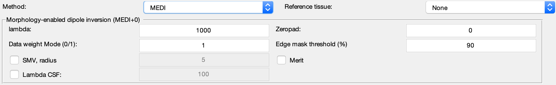
Supported algorithms in SEPIA

  • Phase Unwarpping Algorithms
  • Background Magnetic Field Removal Algorithms
  • QSM Algorithms
    • How can we map the magnetic susceptibility sources from local field map?
      • Thresholded k-space Division (TKD)
      • Closed-form solution with L2-norm regularisation
      • Nonlinear Dipole Inversion (NDI)
      • Iterative LSQR (iLSQR STI Suite)
      • Iterative LSQR (iLSQR)
      • FAst Nonlinear Susceptibility Inversion (FANSI)
      • STreaking Artifact Reduction for QSM (Star-QSM)
      • Morphology enabled dipole inversion (MEDI)
      • MRI Susceptibility Calculation Methods
      • QSMnet+
      • LP-CNN
      • xQSM
  • SWI/SMWI Algorithms
  • R2* Algorithms
  • Subcortical structure segmentation in SEPIA
  • Weightings in SEPIA
  • Lookup table of algorithm parameters

Demonstration

  • Choosing a right phase unwrapping method for your data

Tutorial

  • SEPIA 101 - First QSM with SEPIA
  • Integration of New Phase unwrapping/BFR/QSM Method in SEPIA: Part 1
  • Integration of New Phase unwrapping/BFR/QSM Method in SEPIA: Part 2
  • (f)MRI Toolkit 2019

Acknowledgements and References

  • Acknowledgments
  • References
SEPIA
  • Docs »
  • QSM Algorithms »
  • Morphology enabled dipole inversion (MEDI)
  • Edit on GitHub

Morphology enabled dipole inversion (MEDI)ΒΆ

References: Liu, T., Liu, J., Rochefort, L. de, Spincemaille, P., Khalidov, I., Ledoux, J.R., Wang, Y., 2011. Morphology enabled dipole inversion (MEDI) from a single-angle acquisition: Comparison with COSMOS in human brain imaging. Magnetic resonance in medicine 66, 777–783.

LLiu, J., Liu, T., Rochefort, L. de, Ledoux, J., Khalidov, I., Chen, W., Tsiouris, A.J., Wisnieff, C., Spincemaille, P., Prince, M.R., Wang, Y., 2012. Morphology enabled dipole inversion for quantitative susceptibility mapping using structural consistency between the magnitude image and the susceptibility map. Neuroimage 59, 2560–2568.

Liu, Z., Spincemaille, P., Yao, Y., Zhang, Y., Wang, Y., 2018. MEDI+0: Morphology enabled dipole inversion with automatic uniform cerebrospinal fluid zero reference for quantitative susceptibility mapping. Magnetic resonance in medicine 79, 2795–2803.

../../_images/MEDI.png
Next Previous

© Copyright 2019, Kwok-Shing Chan Revision c303530b.

Built with Sphinx using a theme provided by Read the Docs.